Friday, July 24, 2015

Summer Week 9

This week in lab was spent learning to make microscope tape mounts. This is another, superior way of staining fungal cultures for investigation under the microscope.

When making a normal wet mount slide from a culture grown in a dish a researcher will scrape a small sample off the colony. Unfortunately this distorts the morphology of the mycelium, and bunches the sample into a mass, making observations fuzzy at best.

With tape mounts this frustration is eliminated. Instead of scraping the colony, a strip of tape is gently pressed against the top of the petri plate, preserving the shape of the specimen.

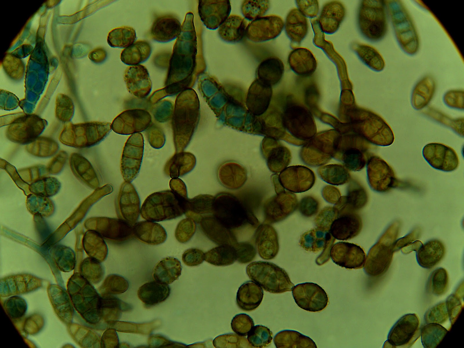
Here are some photos of a colony that was sporulating:


Thursday, July 23, 2015

Summer Week 8

For the first time in my life I extracted DNA! Naturally, I started with one of the most notoriously difficult organisms to extract DNA from- fungi. Fungi are similar to plants in that their structure and support comes from rigid cell walls. However, in fungi the macro-molecule that the cell wall is made of is usually chitin instead of cellulose.

These macro-molecules must be broken down to extract any DNA, which is difficult because of how tough they are. Broadly, there are two ways to break down the cell walls; chemical or mechanical. Chemical degradation of the cell walls would be things like adding enzymes whereas mechanical is more grinding of samples or vortexing with glass beads to shred the walls. The method I used did a little of both.

To make grinding easier and more productive I flash froze mycelium samples in a bath of super cooled isopropyl alcohol. This makes the cytoplasm freeze and the walls brittle so grinding with a pestle is easier. After that I used an E.Z.N.A. Fungal DNA extraction kit that was full of buffers to further degrade the cell walls. I ran a gel on my resulting DNA, popped it in the UV viewer, and this is what I got:


Thursday, July 9, 2015

Summer Week 7

This week in lab I did a variety of things.

I've noticed in the past that the agar in my petri plates will dry out and crack, a process called desiccation. To prolong the life of my endophytes, I have decided to move cultures to new plates. Because I no longer need to select against any bacteria or fast growing fungi I have decided to move my cultures to plates filled with straight potato dextrose agar, no antibiotics or rose bengal dye. So on Monday I made PDA and filled my plates with it.

On Tuesday I moved my cultures. I have also noticed that some of my plates will grow a black fungal contaminate which I assume is coming from the air. To prevent this from happening again I used the laminar hood when moving my cultures. Hopefully by sterilizing the air with UV light there will not be any contamination on my newest plates.

Tuesday, July 7, 2015

Summer Week 6

This week in lab I spent a lot of time staining the fungi that I've grown to identify morphology and hopefully the species.

I have been using lactophenol cotton blue dye in accordance with a protocol from Leck (1999). It is a relatively simple protocol, you basically just add dye to a sample on a slide, but I was really struggling with it. The samples of mycelium I was taking from my plates were too small at first, then they were too thick and only a small portion of the sample absorbed dye.

Finally, after much trail and error, I was able to get a sample clear enough to see structures. The trick was to use dissecting needles to tease out the mycelial mat and wait five min for the dye to absorb into the cell walls. I also discovered that the commassie blue dye works about as well as the lactophenol cotton blue and is a less toxic compound, so I may begin to use that more.


Preparation of Lactophenol Cotton Blue Slide Mounts:
http://www.ncbi.nlm.nih.gov/pmc/articles/PMC1706009/