Thursday, November 19, 2015

Fall 2015 Week 11

This week in lab I haven't done much on my endophyte project. I transferred very small squares of mycelium from the original test plates to plates that have fewer biological controls so I can characterize them.

The rest of the week I worked with Matt on the allelopathy project we have been doing. Some plants have compounds in their leaves that, when they fall to the ground, prevent seeds in the neighboring area to not germinate to lower the competition for resources. We are composting leaves that we know to have allelopathic traits and seeing if the chemicals prevent various seeds to germinate. So I spent all week counting seeds into petri plates in preparation of this experiment.

Friday, November 13, 2015

Fall 2015 Week 10

I ran my last sterilization experiment of the semester this week in lab. The grass plates I made weeks ago are as sterile as the day I put them in my cabinet and so I decided to go ahead with the procedure I used on the grass. This was a summary of my procedure:

  1. Rinse leaves in running tap water for 5 minutes to remove any debris or dirt
  2. Soak leaves in 75% ethanol for 30 seconds
  3. Soak leaves in 4% bleach for 5 minutes
  4. Soak leaves in 75% ethanol for 30 seconds to rinse any residual bleach from the leaves
  5. Cut the leaves in to small squares and put pieces in a petri plate 

This time I varied the times I soaked the silver leaved nightshade leaves in bleach to see if it had any affect. I soaked the leaves in bleach for 5 minutes, 6 minutes, 8 minutes, 10 minutes, and 15 minutes. Things began to grow on most of the plates by Thursday and by today the growth was dense enough to see without squinting. WHAT DOES THIS MEAN? I have no clue, Cori has no clue, Matt has no clue. So I'm just going to characterize this fungi for my project. Its everywhere on these plants.

To characterize the fungi I will use their distinctive characteristics when growing on petri plates, lumpy green with white mycelium on the fringes:

What the mycelium looks like stained and put under a microscope:

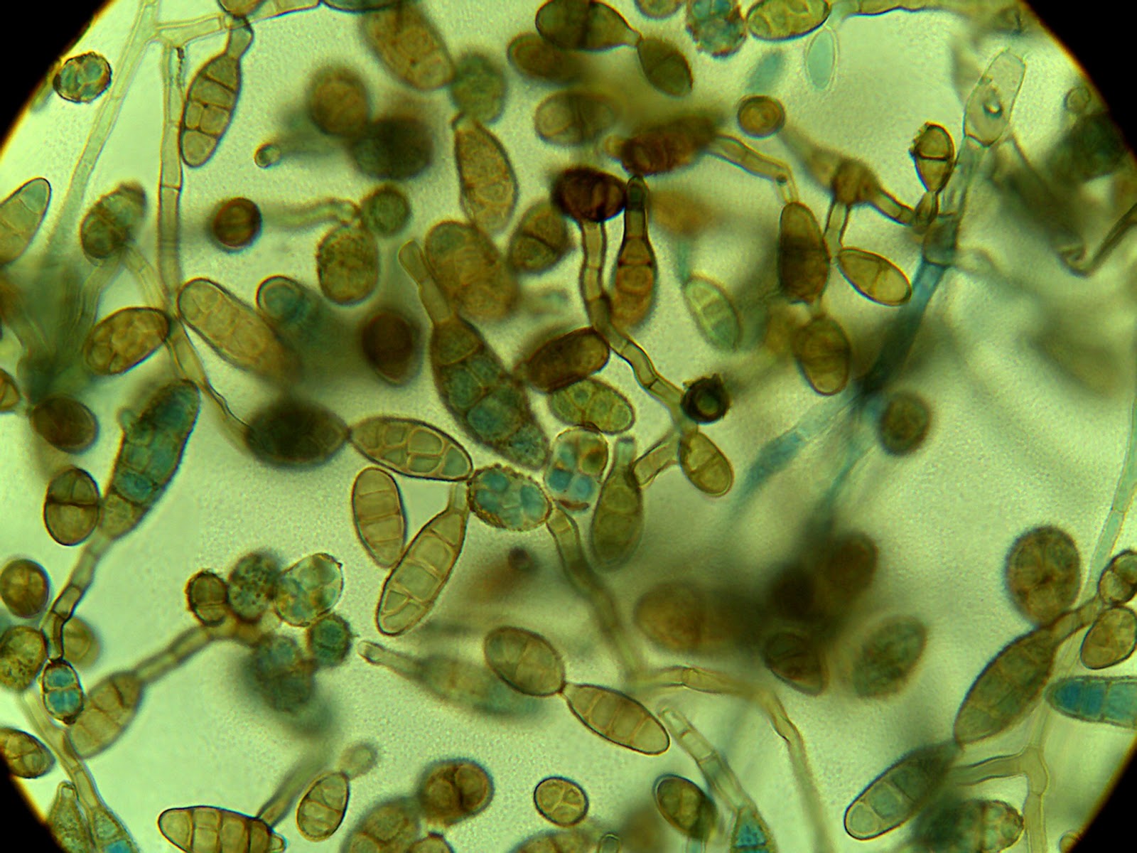
And what the spores look like under a microscope: 

I will also look up any biochemical tests I could possibly do to narrow the search to a genus of fungi I'm working with. 


Thursday, November 5, 2015

Fall 2015 Week 9

This week in lab there has been no growth on my grass species plates, indicating the sterilization procedure is working for the grass. Growth of the gray mold is significantly quicker than the week I've given it, with noticeable mycelium by day 3 or 4. Next week I plan on using the same concentrations of alcohol and bleach but letting the silver leaved nightshade leaves sit longer than 5 minutes so the bleach penetrates the trichomes.

While waiting to see results from my grass plates I have been working more with my unknown bacteria. To even begin to classify a bacterium species one must know the cell shape, whether it is Gram positive or negative, and the organism's oxygen requirements. The first two pieces of information can be deciphered using Gram staining but the last is a requires other tests.

To test if a bacterium is capable of growing in oxygen free environments, two test chambers are prepared. The first is in a giant pickle jar with a burning candle that will use up the oxygen in the container. Only a very small amount of oxygen can get in through the edges of the lid making an environment most suitable to microaerophiles: bacteria that like a very specific, low level of oxygen.

The other test is a box with a pouch of chemicals that use up the all the oxygen in the box, while producing carbon dioxide. This creates an environment that only strict anaerobes or facultative anaerobes can live in. Strict anaerobes are bacteria that can only live in oxygen free environments, which I know I don't have because my bacteria was growing fine on my plates in open air. Facultative anaerobes are bacteria that can switch from oxygen rich environments to oxygen poor environments. 

And as a bonus a photo of my bacterium Gram stain under the microscope:


Thursday, October 29, 2015

Fall 2015 Week 8

This week was the club Halloween fundraiser thingy so I wasn't in lab much.
On Tuesday 10/27 I did do one sterilization experiment, using the endophyte infected grass I've been growing in the plant incubator. The sterilization procedure I used for my last experiment was formulated specifically for grass species, and if I know for sure that my grass has been grown from endophyte infected seeds, then I should be able to grown something besides gray mold.

When looking at the silver leaved nightshade under the microscope there are dense trichomes on the surface of the leaf. Trichomes are fine growths on the surface of many plants that can serve many functions. They look like silver hairs protecting delicate plant tissue from harsh sunlight. They can also help prevent loss of water by creating a small bubble of moister air near the surface of the leaf. This is clearly on advantage in the desert where silver leaved night shade lives. Incidentially it is the dense mat of trichomes on SNS that gives it its name. Here is a photo of the SNS leaves under the microscope. The triangular snowflake structures are the trichomes.


I figure If I can get something besides contamination to grow on my grass I know that the procedure works and I simply need to let the silver leaved night shade soak in bleach a little longer to get it to penetrate between the trichomes of the leaf.

Thursday, October 22, 2015

Fall 2015 Week 7

Some of the literature about endophytes I've read in the past have said more diversity can be found if plant metabolites are on the petri plates. So a few weeks ago I ran an experiment where I blended up some S. eleaegnifolium leaves, strained the slurry and poured the liquid on petri plates filled with PDA, rose bengal dye and streptomycin to see if I could grow anything other than the usual fungi.

Unfortunately I forgot to surface sterilize the leaves I blended up but I did grow something really interesting. On my plates were small, rounded, sticky looking, creamy white colonies that could have been either bacteria or yeast. I isolated it to a few different media but it only grew well on the TSA plate. Here's what it looked like after a few days: 


But it's way cooler now: 

I did a gram stain and found out the organism is a gram positive bacillus bacterium. This is especially interesting because the streptomycin on the plates I make is supposedly broad spectrum enough to kill most bacteria. The plates were made in April so maybe the antibiotics degrade, I thought. I made a streak plate of 4 different bacteria species I knew to be susceptible to streptomycin on one of the last plates and nothing grew. I assumed both that my antibiotics were working and the bacterium was immune to streptomycin. Then I did an antibiotic test plate with four different antibiotic disks: Streptomycin, vancomycin, penicillin and chloraphenicol. Here's what I found:

Everything except penicillin has a zone of inhibition. WHAT? Not what I expected. The concentration of the antibiotic disk was 10 micrograms, about what the concentration in the agar should have been. I looked back in my notebook and guess what? I diluted my streptomycin to about 1/10 of the strength I thought it was. Decimals are hard to keep track of. So now I know those plates were not exactly what I thought but they did their job well enough while I used them. Interesting. 

Thursday, October 15, 2015

Fall 2015 Week 6

This week in lab I ran another sterilization experiment. This time I followed a protocol exactly. I soaked the nightshade leaves in 75% ethanol for three min then in 4% bleach for 5 minutes then ethanol again for 45 seconds. I made nine plates, the more specimens the more data to look at, and have begun to keep careful track of the how things grow on the plates using an Excel spread sheet.

I've found that, once again, within a few days I'm seeing significant fungal growth on all my plates. Its early to know for sure, but I'm pretty sure its the same green-grey mold stuff I've been growing all semester. I'm going to try a few more experiments to attempt to isolate the source of the contamination. I will try plating the tap water I'm rinsing the leaves in as well as test my glassware. I will also think about increasing either the concentration of the bleach or the amount of time spent soaking in the bleach solution.

Another thing I want to try is sterilizing the grass I'm growing that, according to the package, has endophytes in the leaves. If I can isolate endophytes from there I know I'm on the right track.

This is a picture of all the plates I made Spring 2015. They are arranged by decreasing times of sterilization, the first row being 10 minutes and the last being unsterilized. This photo shows that many different kinds of fungi are growing on unsterilized leaves and almost nothing is growing on the longer sterilization times.


Thursday, October 8, 2015

Fall 2015 Week 5

This week in lab I did a lot of little tasks to maintain my experiments. I made more media (I simultaneously feel that I don't use my media fast enough and that I spend all my time making media), I replanted endophyte infected grass seeds, took some photos off the lab camera and diluted alcohol (though I now realize I should have made more bleach too).

The plates I made last week testing the sterility of different bleach concentrations were over run with contamination as well. All of the plates had the same green/black mold as before, no matter how high the bleach concentration. I'm beginning to feel frustrated.

Next week I plan on using a sterilization technique I found using 75% ethyl-alcohol and 4% bleach solution-way stronger than any I've used before. They say to dip the plant tissue in alcohol, then bleach, then alcohol again. Apparently the second alcohol dip removes some of the bleach residue, which could be one reason my original experiment with 1.25% bleach did not grow anything at longer sterilization times.

Contamination or not?