How time flies! School has been in session for nearly three weeks, all my classes are now in full swing and I'm finally getting back into the rhythm of things.
Most of this week has been spent catching up with all the lab people and getting to know some new faces. Other than that I've been using the light microscope to try and find endophytes in silver leaved night shade leaves before sterilizing and plating them. It hasn't been as easy as during the summer because the health status of the individuals I'm taking samples from keeps changing. First I thought they were all completely dried out and dead but, now my brother has started to water the front lawn, where these plants grow, there are tons of healthy, unstressed plants without endophytes. I feel like I need to control their growing conditions more stringently.
Here is a photo of Silver leaved nightshade or Solanum elaeagnifolium:
Saturday, September 12, 2015
Tuesday, August 11, 2015
Summer Week 12
This week I have not been in lab. I haven't even been in Phoenix for over a week. Hurray!!!
Instead of sitting in a boring, stuffy lab all day (haha not really), I have been running all around Colorado. My friends Luke and Jason moved here over two years ago and this is the first time I've seen them since then. Its been so great getting caught up with them and adventuring around Colorado.
I'm looking forward to getting back in the lab when school starts again. For now, here is a photo of beautiful Boulder:
Instead of sitting in a boring, stuffy lab all day (haha not really), I have been running all around Colorado. My friends Luke and Jason moved here over two years ago and this is the first time I've seen them since then. Its been so great getting caught up with them and adventuring around Colorado.
I'm looking forward to getting back in the lab when school starts again. For now, here is a photo of beautiful Boulder:
Sunday, August 2, 2015
Summer Week 11
I'm doing my blog early this week because I'm headed to Denver WOOT!
I anticipate most of my time will be spent working on my final project. Thankfully the structure of my project is similar to the one I did in the Spring, so much of the background information and literature citations do not need to be reexamined.
One thing that I've been more concerned about since looking at my past research is my sterilization method. Most of the literature concerning endophytes says to use a low concentration of sodium hypochlorate, bleach, to sterilize the surface of plant tissues. I, however, found that the bleach was so caustic that it killed all possible endophytes.
In this table from Spring 2015 shows what kinds of fungi I grew based on how long I soaked the tissues in bleach. C means contaminate, + means possible endophyte and - means no growth at all.
As can be seen after even one minute of soaking in bleach nothing is growing on my plates. There are two possible explanations for this: the bleach solution used last semester was incorrectly diluted making it more concentrated than expected, or bleach is caustic to fungi at any concentration.
I have been getting better results using 100% ethanol for 5 minutes but have only found one other paper that uses exclusively ethanol, which makes me wary...
I anticipate most of my time will be spent working on my final project. Thankfully the structure of my project is similar to the one I did in the Spring, so much of the background information and literature citations do not need to be reexamined.
One thing that I've been more concerned about since looking at my past research is my sterilization method. Most of the literature concerning endophytes says to use a low concentration of sodium hypochlorate, bleach, to sterilize the surface of plant tissues. I, however, found that the bleach was so caustic that it killed all possible endophytes.
In this table from Spring 2015 shows what kinds of fungi I grew based on how long I soaked the tissues in bleach. C means contaminate, + means possible endophyte and - means no growth at all.
As can be seen after even one minute of soaking in bleach nothing is growing on my plates. There are two possible explanations for this: the bleach solution used last semester was incorrectly diluted making it more concentrated than expected, or bleach is caustic to fungi at any concentration.
I have been getting better results using 100% ethanol for 5 minutes but have only found one other paper that uses exclusively ethanol, which makes me wary...
Saturday, August 1, 2015
Summer Week 10
This week in lab has been spent in contemplation of my summer. As it nears to an end, and I begin to write my final paper, I am compelled to think about all I have accomplished:
- Staining plant tissues to identify endophytes microscopically
- Perfecting my sterilization technique to include the laminar hood
- Moving my fungi from bengal agar to potato dextrose agar
- Using tape mounts to stain fungal structures
- Using super cooled ethanol to freeze dry fungal tissues
- Extracting DNA with a kit
I certainly have had a productive summer. But I think the most important thing I did this summer was sit down and discuss with Cori how to organize my data. I have been struggling with this since I got into this program. What to write down and in what format. Cori really helped me understand how it is people in labs organize data and helped me make more sense of my lab notebook. She's a great mentor
This is a video showing my freeze drying setup
Friday, July 24, 2015
Summer Week 9
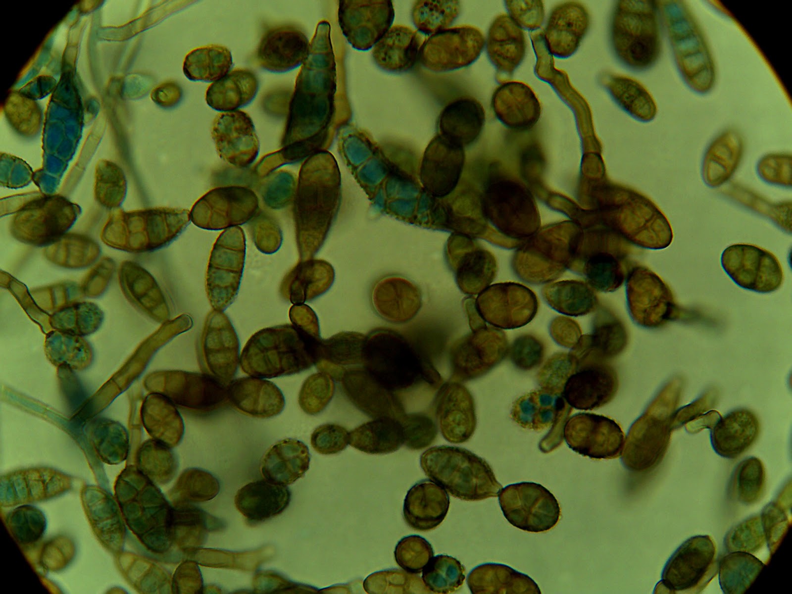
This week in lab was spent learning to make microscope tape mounts. This is another, superior way of staining fungal cultures for investigation under the microscope.
When making a normal wet mount slide from a culture grown in a dish a researcher will scrape a small sample off the colony. Unfortunately this distorts the morphology of the mycelium, and bunches the sample into a mass, making observations fuzzy at best.
With tape mounts this frustration is eliminated. Instead of scraping the colony, a strip of tape is gently pressed against the top of the petri plate, preserving the shape of the specimen.
Here are some photos of a colony that was sporulating:
When making a normal wet mount slide from a culture grown in a dish a researcher will scrape a small sample off the colony. Unfortunately this distorts the morphology of the mycelium, and bunches the sample into a mass, making observations fuzzy at best.
With tape mounts this frustration is eliminated. Instead of scraping the colony, a strip of tape is gently pressed against the top of the petri plate, preserving the shape of the specimen.
Here are some photos of a colony that was sporulating:
Thursday, July 23, 2015
Summer Week 8
For the first time in my life I extracted DNA! Naturally, I started with one of the most notoriously difficult organisms to extract DNA from- fungi. Fungi are similar to plants in that their structure and support comes from rigid cell walls. However, in fungi the macro-molecule that the cell wall is made of is usually chitin instead of cellulose.
These macro-molecules must be broken down to extract any DNA, which is difficult because of how tough they are. Broadly, there are two ways to break down the cell walls; chemical or mechanical. Chemical degradation of the cell walls would be things like adding enzymes whereas mechanical is more grinding of samples or vortexing with glass beads to shred the walls. The method I used did a little of both.
To make grinding easier and more productive I flash froze mycelium samples in a bath of super cooled isopropyl alcohol. This makes the cytoplasm freeze and the walls brittle so grinding with a pestle is easier. After that I used an E.Z.N.A. Fungal DNA extraction kit that was full of buffers to further degrade the cell walls. I ran a gel on my resulting DNA, popped it in the UV viewer, and this is what I got:
These macro-molecules must be broken down to extract any DNA, which is difficult because of how tough they are. Broadly, there are two ways to break down the cell walls; chemical or mechanical. Chemical degradation of the cell walls would be things like adding enzymes whereas mechanical is more grinding of samples or vortexing with glass beads to shred the walls. The method I used did a little of both.
To make grinding easier and more productive I flash froze mycelium samples in a bath of super cooled isopropyl alcohol. This makes the cytoplasm freeze and the walls brittle so grinding with a pestle is easier. After that I used an E.Z.N.A. Fungal DNA extraction kit that was full of buffers to further degrade the cell walls. I ran a gel on my resulting DNA, popped it in the UV viewer, and this is what I got:
Thursday, July 9, 2015
Summer Week 7
This week in lab I did a variety of things.
I've noticed in the past that the agar in my petri plates will dry out and crack, a process called desiccation. To prolong the life of my endophytes, I have decided to move cultures to new plates. Because I no longer need to select against any bacteria or fast growing fungi I have decided to move my cultures to plates filled with straight potato dextrose agar, no antibiotics or rose bengal dye. So on Monday I made PDA and filled my plates with it.
On Tuesday I moved my cultures. I have also noticed that some of my plates will grow a black fungal contaminate which I assume is coming from the air. To prevent this from happening again I used the laminar hood when moving my cultures. Hopefully by sterilizing the air with UV light there will not be any contamination on my newest plates.
I've noticed in the past that the agar in my petri plates will dry out and crack, a process called desiccation. To prolong the life of my endophytes, I have decided to move cultures to new plates. Because I no longer need to select against any bacteria or fast growing fungi I have decided to move my cultures to plates filled with straight potato dextrose agar, no antibiotics or rose bengal dye. So on Monday I made PDA and filled my plates with it.
On Tuesday I moved my cultures. I have also noticed that some of my plates will grow a black fungal contaminate which I assume is coming from the air. To prevent this from happening again I used the laminar hood when moving my cultures. Hopefully by sterilizing the air with UV light there will not be any contamination on my newest plates.
Subscribe to:
Posts (Atom)